• Professional healthcare
  • Products & Services
  • Inspiration
  • Support & Contact
  • Shop
‌
‌
‌
‌
‌
‌
‌
‌
‌
‌
‌
‌
‌
‌
‌
‌
‌
‌
‌
‌
  • Consumer productsConsumer products
    • Home
    • Support
    • Product registration
    • My Philips
  • Healthcare professionalsHealthcare professionals
    • Home
    • Products
    • Services
    • Specialties
    • Solutions
  • Other business solutionsOther business solutions
    • Lighting
    • Automotive
    • Dictation Solutions
    • More
    • Hearing solutions
    • Display solutions
  • About usAbout us
    • Home
    • News
    • Investor Relations
    • Careers
    • More
    • Innovation
    • Environmental, Social and Governance
  • Contact and supportContact and support
    • Customer support
    • Professional healthcare support
    • Company contacts
  • Stay up-to-dateStay up-to-date
  • Select your country / languageGlobal / English
  • Privacy Notice
  • Terms of use
  • Cookie notice
© Koninklijke Philips N.V., 2004 - 2025. Alla rättigheter förbehålles.
‌
‌
‌
‌
‌
‌
‌
‌
‌
‌
‌
‌
‌
‌
‌
‌
‌
‌
‌
‌
  1. Diagnostic imaging
  2. Magnetic resonance imaging
  3. MRI software
  4. MRCAT Head and Neck
MRCAT Head and Neck
MR-RT clinical application
MRI software
‌
‌
‌
‌
‌
‌
‌
‌
‌

MRCAT Head and Neck

MR-RT clinical application
MRI software
MRCAT Head and Neck enables the use of MRI as a primary imaging modality for radiotherapy planning of patients with soft tissue tumors in the Head and Neck, without the need for CT. Powered by AI, MRCAT (MR for Calculating ATtenuation) images with CT-like density information are automatically generated from a single, high-resolution MR scan and can be used for CT-equivalent¹ dose calculations and accurate² patient positioning.

All Philips professional healthcare customers can open, track and manage equipment service tickets in our Customer Services Portal at no cost. Register for an account here.
For all support resources and contact information, visit our Support hub.

Technical Data Sheet
Technical data sheet(301.11 KB)
See all
MR-only sim for soft tissue tumors in the head and neck region
Short scan times promote patient comfort
Automatic generation of CT-like density information using AI

Clinical image gallery

  • MRCAT-based and CT-based dose plans
    MRCAT-based and CT-based dose plans
  • MRCAT Head and Neck with continuous Hounsfield Units
    MRCAT Head and Neck with continuous Hounsfield Units
  • MRCAT generation based on single mDIXON source scan
    MRCAT generation based on single mDIXON source scan
  • MRCAT-based patient positioning
    MRCAT-based patient positioning
Features
Unleash the real power of MR simulation
MRCAT Head and Neck lets you plan radiation therapy using MRI only, without the need for CT. Within just one fast MR exam, MRCAT Head and Neck provides excellent soft-tissue contrast for target and OAR delineation, and CT-like density information for dose calculations. This not only extends the benefits of MRI’s excellent soft-tissue contrast to radiotherapy planning and adaptive strategies, but it also eliminates arduous, error-prone CT-MRI registration from the process.
MR Simulation
Consistent, accurate imaging protocol
Image acquisition is made easy by a dedicated ExamCard that includes a single, multi-contrast 3D T1W mDIXON scan, which is standardized to deliver consistent results and high geometric accuracy. The MRCAT Head and Neck scan can be acquired before or after contrast agent administration. Additional sequences, e.g. for delineation, can be easily added to the protocol to address your specific clinical needs.
Protocol
Short scan times promote patient comfort
Head and neck cancer patients often find it difficult to endure an MRI exam, especially when immobilized by a thermoplastic mask. The high-resolution 3D MRCAT source scan is accelerated by Compressed SENSE and requires < 3 minutes to complete – while covering a large FOV (up to 380 mm in the feet-head direction). This promotes patient comfort, as well as productivity by minimizing time in the scanner.
Patient Comfort


Specifications
  • MRCAT Head and Neck
    Compatibility MR system
    • Ingenia MR-RT 1.5T and 3.0T
    • Ingenia Ambition MR-RT 1.5T S and X
    • Ingenia Elition MR-RT 3.0T S and X
    • Ingenia MR-RT Evolution 1.5T and 3.0T

Documentation

Technical data sheet
PDF|301.11 KB

Recommended products

  • Ingenia MR-RT XD Versatility to fit your planning
    Ingenia MR-RT XD
    The Philips Ingenia MR-RT XD platform harnesses the power and value of MRI for radiation therapy planning. It has been designed around the needs of radiation oncology, with ease-of-use, streamlined integration, and versatility in mind. Central to that concept is the ability to define a tailored approach with customizable functionality that meets your individual clinical, workflow, and budgetary requirements – all to provide better patient care.
  • MRCAT Brain MR-RT clinical application
    MRCAT Brain
    MRCAT Brain clinical application allows the use of MRI as the primary imaging modality for radiotherapy planning of primary and metastatic tumors in the brain without the need for CT. Detailed anatomical information for contouring and attenuation maps for dose calculations are both obtained from a single, submillimeter resolution 3D T1W mDIXON MR sequence. Artificial Intelligence (AI) is used for fast computation of continuous Hounsfield units directly on the MR console.
  • MRCAT Prostate + Auto-Contouring MR-RT clinical application
    MRCAT Prostate + Auto-Contouring
    As a plug-in clinical application to Ingenia MR-RT, MRCAT Prostate + Auto-Contouring provides attenuation maps and automated, MR-based contours of prostate and organs at risk in as little as 20 minutes – all in a repeatable ‘one-click’ workflow.
  • MRCAT Pelvis MR-RT clinical application
    MRCAT Pelvis
    MRCAT Pelvis lets you plan radiation therapy using MRI as a single modality solution. Within just one MR exam, MRCAT Pelvis provides excellent soft-tissue contrast for target and OAR delineation, and continuous Hounsfield units for dose calculations. MRCAT (MR for Calculating ATtenuation) data can be used for export to treatment planning systems for CT-equivalent** dose calculations. In addition, MR-based imaging enables CBCT-based positioning based on soft-tissue contrast with the look and feel of CT.
Disclaimer
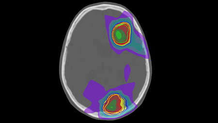
¹ The mean dose in the PTV does not differ more than 1% in MRCAT-based plans as compared to CT-based plans for 95% of the patient cases
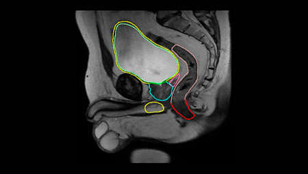
² Accurate means: MRCAT-based patient positioning is within 2 mm accuracy for bony anatomy compared to CT-based patient positioning for 95% of cases
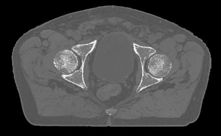
³ Accurate means: MRCAT provides < ± 1 mm total system geometric accuracy of image data in < 20 cm Diameter Spherical Volume (DSV) and < ± 2 mm total system geometric accuracy of image data in < 40 cm Diameter Spherical Volume (DSV)* * Limited to 32 cm in z-direction in more than 95% of the points within the volume
MRCAT Head and Neck is not available for sale in all markets.