Artifacts from large metal objects, such as orthopedic implants, can make it difficult and time-consuming to generate contours of critical structures and target volumes. The Philips metal artifact reduction algorithm for orthopedic implants, O-MAR, improves image quality and visualization of anatomy by reducing the effect of metal on the images. This helps speed up workflow in simulation and treatment planning when metal orthopedic implants are present. This material is not for distribution/use in the USA
Fast and accurate contouring for patients with orthopedic implants
Metal artifacts can be visible as streaks and dark areas in CT images. When CT is used for radiation treatment planning, it is vital that the tumor and organs at risk be accurately identified and delineated. Orthopedic implants can become a critical impediment: the image artifacts they cause may have to be manually compensated for in the plan. The O-MAR algorithm helps you provide fast and accurate treatment planning for patients with orthopedic implants. It isolates the effects of large metal objects in the image data and improves image quality. Users switch on O-MAR by simply clicking a check box on the image acquisition console of their Philips CT system. The algorithm automatically produces conventional images and corrected images for clinician review.
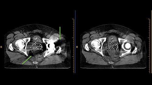
Metal artifacts in CT images, manifesting as dark streaks (green arrows). O-MAR enhances the visualization of structures by iteratively calculating interpolated values which simulate tissue.
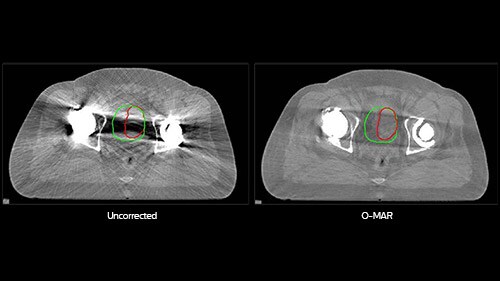
Bladder contour: O-MAR contour in red. Uncorrected contour in green. Volume difference: 32%. On the uncorrected images, the bladder was mostly obscured by the dark shadow caused by metal, so the physician overestimated the size of the bladder. On the O-MAR images, the size of the bladder was visible; there was no inter-observer variability.
Improving image quality, one step at a time
O-MAR is an iterative process that improves the image step by step. First, O-MAR identifies which pixels in the image belong to the orthopedic implant. If no sufficiently large clusters of metal pixels are present in the image, no further processing is performed – O-MAR has no effect on non-metal images and is not applied to stents or similar small metal objects. Second, the algorithm enters its iterative loop. Each repetition of the process subtracts a correction image from the input image. The resulting image can then become the new input image, refining the results. Though O-MAR does not totally eliminate metal artifacts, it can reduce their effect on CT images to significantly enhance their diagnostic quality.
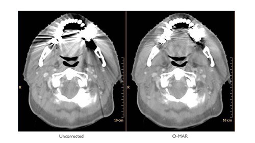
O-MAR helps you see in the dark areas
With O-MAR, not only are severe streaking artifacts reduced, substantial portions of previously obscured anatomy can now be visualized. Since the system will always reconstruct both sets of images whenever O-MAR is selected, the uncorrected images are readily available. Although the main purpose of O-MAR is to address artifacts arising from orthopedic metal, it is also effective for some kinds of non-orthopedic metal, e.g. dental fillings.