
maj 20, 2019
Pioneering new augmented-reality surgical navigation technology for precision surgery
In spine surgery, one of the biggest challenges is to place implants in the vertebrae for the treatment of patients with spinal malformations, fractures, and other back conditions. The margin of error can be as small as 1 millimeter, because the surgical area is very close to the spinal cord, arteries and other vital structures. To help surgeons navigate through these delicate areas, Karolinska University Hospital is pioneering the use of a new augmented-reality surgical navigation technology developed in collaboration with Philips, as part of a wider innovation partnership.
Dr. Adrian Elmi Terander, associate professor and senior consult neurosurgeon at Karolinska, sees the joint innovation project holding great promise. Augumented Reality and intraoperative imaging enables us to place spinal implants much safer and more accurate.
Augumented Reality and intraoperative imaging enables us to place spinal implants much safer and more accurate.
Dr. Adrian Elmi Terander
Neurosurgeon at Karolinska University Hospital
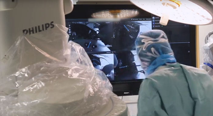
Real-time imaging and navigation support
Surgical navigation systems are not new – they have been used for the past 20-30 years. Traditionally in spine surgery, X-rays of a patient are taken before the operation. These scans are then used to plan and guide the operation. With the new navigation technology, the care team gets real-time imaging and real-time navigation support during the operation, both in one system. A flat detector with four integrated hi-resolution cameras captures external images of the patient on the operating table. These images are automatically combined with internal images of the bony anatomy captured by an X-ray system, generating 3D views with augmented reality to help guide the surgical procedure. “This allows us to plan our procedures virtually,” Dr. Elmi Terander says. “We can now insert implants with high precision to improve patient safety.” Indicating some of the benefits of the technology, the first in human clinical study using AR for spine surgery, published in the journal Spine [1] has shown that the technology enables a high (94.1%) overall screw placement accuracy whilst virtually eliminating radiation exposure to the surgeon and OR staff.
Instant post-surgery checks
An additional advantage of the new augmented reality technology is that the result of the surgical procedure can be checked – and if necessary, corrected – in the operating room, directly after the operation. “Traditionally, the patient would go back to the general ward after the operation, and undergo a CT scan the next day,” Dr. Elmi Terander explains. “With this new technology, we can do a 3D check in the operating room, to see if all the implants have been inserted correctly. If we need to make adjustments, we can do this straight away, saving the patient a second operation.”
With this new technology, we can do a 3D check in the operating room, to see if all the implants have been inserted correctly. If we need to make adjustments, we can do this straight away, saving the patient a second operation.
Dr. Adrian Elmi Terander
Neurosurgeon at Karolinska University Hospital
The whole surgical procedure is intended to minimize minimal radiation exposure for the patient and to avoid radiation exposure for the surgeon altogether. “This is also very important for us,” says Dr. Elmi Terander. “Safety of patients and staff is obviously a top priority.”
Breaking boundaries
While working on this innovation project, Dr. Elmi Terander has seen specialists from different disciplines within Karolinska come together. “Over the last years, we have had a very fruitful collaboration with our colleagues in neuroradiology, anesthesia and orthopedics, performing surgery together,” he says. “Neurosurgeons and orthopedic surgeons bring different strengths to the operating room: neurosurgeons excel in high-precision work, whereas orthopedics are highly visual 3D thinkers. With this new surgical navigation technology, we can combine these strengths to achieve better outcomes for patients. I think this project is a prime example of what Karolinska is trying to achieve with its new operating model: breaking internal boundaries to deliver the best possible care.”
Looking forward
As a next step, Karolinska will be working closely with Philips to further develop the new augmented reality technology. Dr. Elmi Terander and his team will also start looking at other possible applications. “So far, we have focused on spinal surgery. But when you’re operating on a brain tumor, for example, you also need to know exactly where you are with your instruments. It’s still early days, but I envision a future in which this kind of technology will be used in all parts of the body that require high-precision navigation.” [1] Pedicle screw placement using augmented reality surgical navigation with intraoperative 3D imaging: a first in-human prospective cohort study SPINE, An International Journal for the study of the spine, September 2018 Learn more about the innovation partnership between Karolinska University Hospital and Philips here, and see more published studies from the partnership here.
Fler relaterade nyheter
-
![Träffa Philips på Vitalis – 23-25 maj]()
maj 12, 2023
-
![Philips donerar 22 hjärtstartare]()
maj 01, 2023
-
![Träffa Philips på SAMTIT 3-5 maj]()
april 12, 2023
-
![Hjärtsäkrat Grannskap hjärtsäkrar Sveriges bostadsområden]()
februari 14, 2023

